¡Tu solución está lista!
Nuestra ayuda de expertos desglosó tu problema en una solución confiable y fácil de entender.
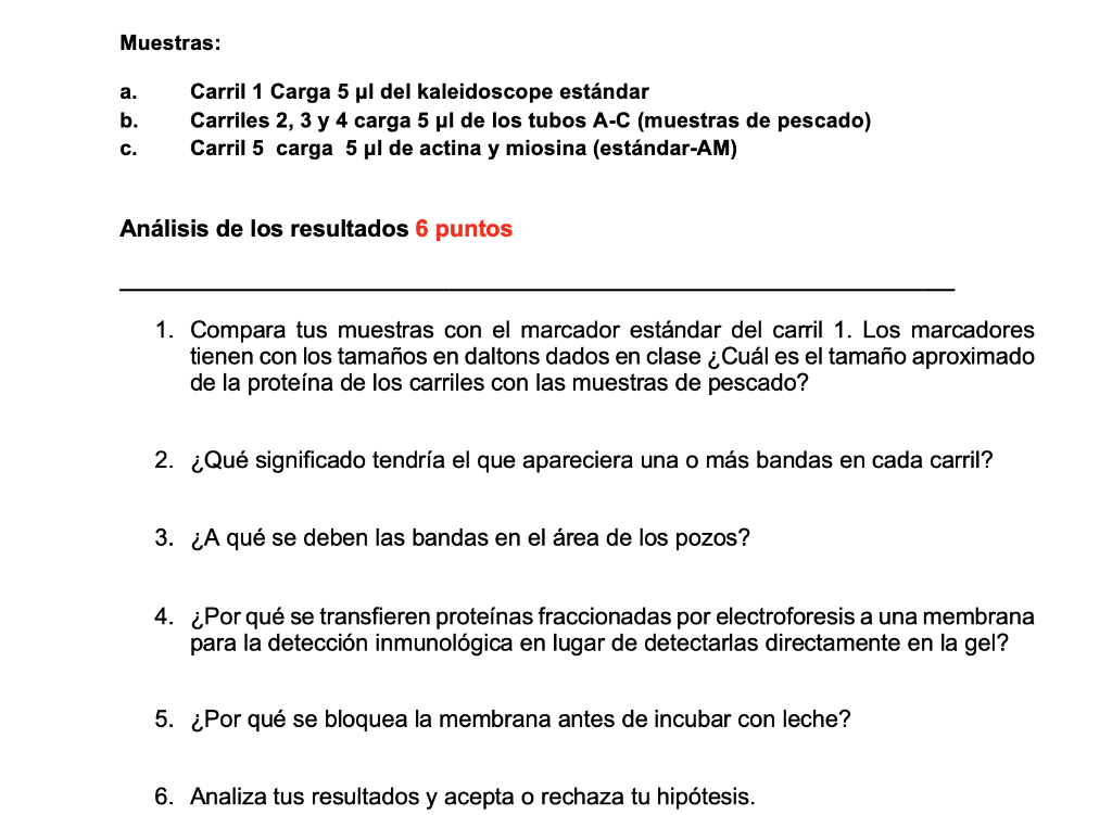
Mira la respuestaMira la respuesta done loadingPregunta: Samples: a. Lane 1 Load 5 μl of the standard kaleidoscope b. Lanes 2, 3 and 4 load 5 μl from tubes A-C (fish samples) c. Lane 5 loads 5 μl of actin and myosin (standard-AM) Analysis of the results 6 points ________________________________________________________________ 1. Compare your samples to the standard marker in lane 1. The markers have the sizes in
Samples: a. Lane 1 Load 5 μl of the standard kaleidoscope b. Lanes 2, 3 and 4 load 5 μl from tubes A-C (fish samples) c. Lane 5 loads 5 μl of actin and myosin (standard-AM) Analysis of the results 6 points ________________________________________________________________ 1. Compare your samples to the standard marker in lane 1. The markers have the sizes in daltons given in class. What is the approximate size of the protein in the lanes with the fish samples? 2. What would it mean if one or more bands appeared in each lane? 3. What are the bands in the area of the wells due to? 4. Why are electrophoretically fractionated proteins transferred to a membrane for immunological detection rather than detected directly on the gel? 5. Why is the membrane blocked before incubating with milk? 6. Analyze your results and accept or reject your hypothesis. 7. According to your results, compare the information you obtained from Fishbase.net of each fish, grouper, snapper and dorado, and determine according to their differences or similarities which of the samples (A, B or C) can be each of the fish studied by comparing the pattern of protein bands on the gel. Include the information of each fish as an annex to the report.
- Esta es la mejor manera de resolver el problema.Solución
2. If more than one band appears in each lane, this means that the antibody which is used for immunological detection of protein binds to more than one protein in a lane. This could possibly result from non -specific binding of antibody to other prot…
Mira la respuesta completa
Texto de la transcripción de la imagen:
Muestras: a. b. Carril 1 Carga 5 pl del kaleidoscope estándar Carriles 2, 3 y 4 carga 5 pl de los tubos A-C (muestras de pescado) Carril 5 carga 5 ul de actina y miosina (estándar-AM) C. Análisis de los resultados 6 puntos 1. Compara tus muestras con el marcador estándar del carril 1. Los marcadores tienen con los tamaños en daltons dados en clase ¿Cuál es el tamaño aproximado de la proteína de los carriles con las muestras de pescado? 2. ¿Qué significado tendría el que apareciera una o más bandas en cada carril? 3. ¿A qué se deben las bandas en el área de los pozos? 4. ¿Por qué se transfieren proteínas fraccionadas por electroforesis a una membrana para la detección inmunológica en lugar de detectarlas directamente en gel? 5. ¿Por qué se bloquea la membrana antes de incubar con leche? 6. Analiza tus resultados y acepta o rechaza tu hipótesis.

Estudia mejor, ¡ahora en español!
Entiende todos los problemas con explicaciones al instante y pasos fáciles de aprender de la mano de expertos reales.
