¡Tu solución está lista!
Nuestra ayuda de expertos desglosó tu problema en una solución confiable y fácil de entender.
Mira la respuestaMira la respuesta done loadingPregunta: Determines the fungus causing the disease A 44-year-old Peruvian patient, resident in Spain, with abdominal pain of one month's evolution. On physical examination, a slightly painful 5 x 10 cm mass was palpated on the right flank, which on ultrasound and opaque enema appeared as an image suggestive of a neoformative and inflammatory process of the ascending
Determines the fungus causing the disease
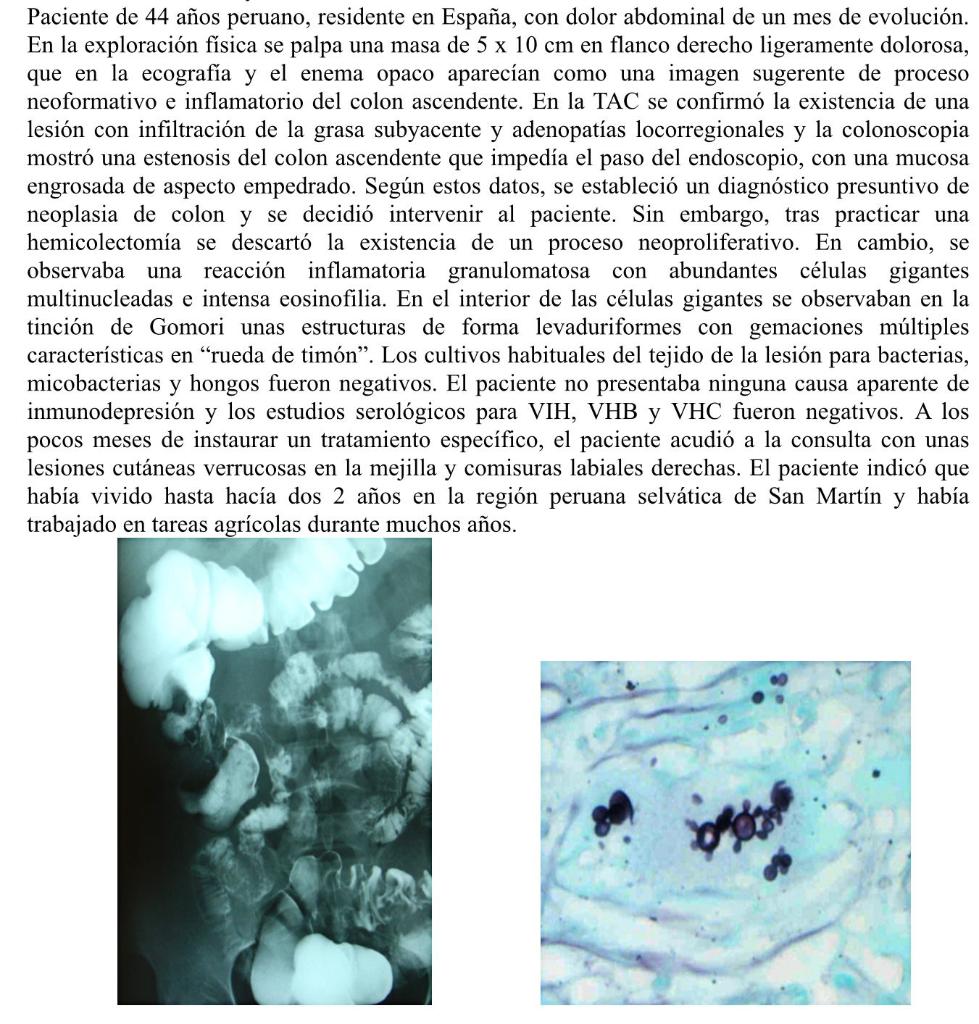
A 44-year-old Peruvian patient, resident in Spain, with abdominal pain of one month's evolution. On physical examination, a slightly painful 5 x 10 cm mass was palpated on the right flank, which on ultrasound and opaque enema appeared as an image suggestive of a neoformative and inflammatory process of the ascending colon. The CT scan confirmed the existence of a lesion with infiltration of the underlying fat and locoregional adenopathies, and the colonoscopy showed a stenosis of the ascending colon that prevented the passage of the endoscope, with a thickened mucosa with a cobbled appearance. Based on these data, a presumptive diagnosis of colon neoplasia was established and it was decided to intervene on the patient. However, after performing a hemicolectomy, the existence of a neoproliferative process was ruled out. Instead, a granulomatous inflammatory reaction with abundant multinucleated giant cells and intense eosinophilia was observed. Within the giant cells, Gomori staining revealed yeast-shaped structures with multiple characteristic “helm wheel” budding. Routine cultures of the lesion tissue for bacteria, mycobacteria, and fungi were negative. The patient had no apparent cause of immunosuppression and serological studies for HIV, HBV, and HCV were negative. A few months after starting a specific treatment, the patient presented with verrucous skin lesions on the cheek and right corners of the lips. The patient indicated that he had lived until 2 years ago in the Peruvian jungle region of San Martín and had worked in agriculture for many years.
 Hay 2 pasos para resolver este problema.SoluciónPaso 1Mira la respuesta completa
The patient's case presents a complex medical scenario, with an initial diagnosis of colon neoplasia...
Paso 2 DesbloqueaRespuestaDesbloquea

Estudia mejor, ¡ahora en español!
Entiende todos los problemas con explicaciones al instante y pasos fáciles de aprender de la mano de expertos reales.